Recurrent Left Abducens Nerve Palsy and Idiopathic Intracranial Hypertension During Pregnancy: A Diagnostic Challenge
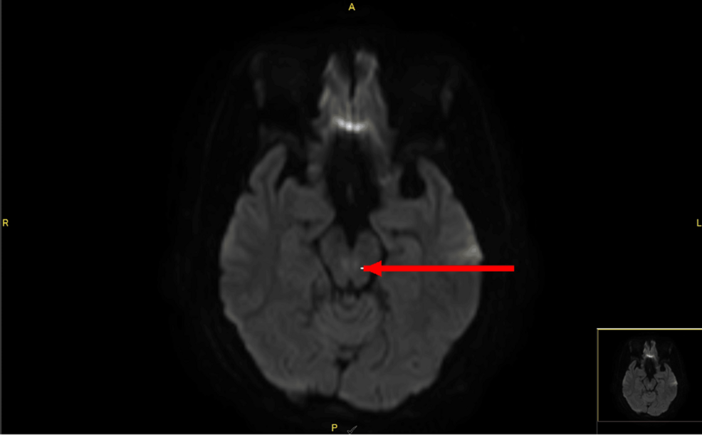
Idiopathic intracranial hypertension (IIH) is characterized by elevated intracranial pressure without an identifiable mass lesion, hydrocephalus, cerebral venous sinus thrombosis, or abnormal cerebrospinal fluid composition. Diagnosis during pregnancy can be challenging because headache and visual symptoms overlap with several neurologic and obstetric disorders. We report a previously healthy 20-year-old primigravida with a body mass index of approximately 33 kg/m² who presented at 17 weeks’ gestation with intermittent headache, photophobia, blurred vision, horizontal diplopia, nasal deviation of the left eye, and absent left-eye abduction. Visual acuity was 6/6 bilaterally, with mild bilateral optic disc swelling, greater on the left. The initial brain magnetic resonance imaging report described subtle paired diffusion-weighted signal abnormalities in the region of the medial longitudinal fasciculi, interpreted as possible mild cytotoxic edema related to a potentially reversible ischemic event. These findings were not anatomically concordant with the documented isolated left abduction deficit, and subsequent informal radiologic review raised the possibility that they were artifactual; no formal radiology addendum was available. Magnetic resonance angiography showed no significant intracranial arterial abnormality, and magnetic resonance venography excluded cerebral venous sinus thrombosis. Lumbar puncture in the lateral decubitus position demonstrated an opening pressure of 31 cmH₂O with normal cerebrospinal fluid constituents, followed by resolution of her symptoms and abducens palsy after drainage. Symptoms recurred at 20 weeks, when repeat opening pressure was 42 cmH₂O; and they again improved after drainage. Acetazolamide was commenced at 250 mg twice daily and increased to 500 mg twice daily at 33 weeks because of persistent optic disc swelling. The patient underwent cesarean delivery at 36 weeks; the specific clinical rationale for the mode of delivery was unavailable in the records reviewed. The neonate weighed 2.6 kg, had Apgar scores of seven and eight, and had no documented neonatal complications. Acetazolamide was continued postpartum. At telephone review approximately six months after delivery, the patient remained clinically well, reported adherence to her prescribed acetazolamide, and denied recurrent headache, blurred vision, diplopia, or ocular deviation. Final objective ophthalmologic resolution could not be confirmed because she did not attend her most recent scheduled ophthalmology follow-up. This case emphasizes the need to integrate clinical examination, cerebrospinal fluid findings, and neuroimaging when evaluating intracranial hypertension during pregnancy.
News providers only return a preview of the article body. Continue reading the full story at the original source.
Cureus →